top of page

3D Illustrations
The following illustrations were created using 3D modelling software


Please Scroll
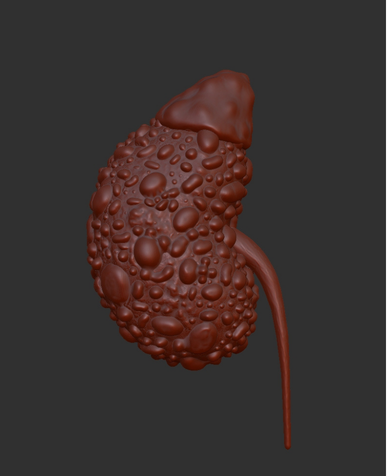
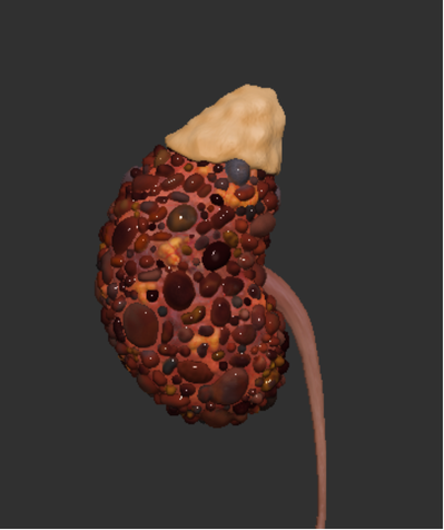
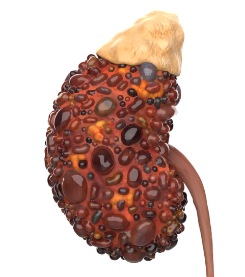
Polycystic kidney disease animation
This 3D model of a polycystic kidney began with sculpting the base in ZBrush , as well as adding colour. The final model, including a healthy kidney and bladder was then exported to KeyShot to add lighting and create the animation.
Following this, the final video was edited using Adobe After Effects to produce a resource aimed to educate the patients and the general population about this disease.
Facial Nerve Course - 3D Interactive Model
This project was created in collaboration with a professor of anatomy at the University of Dundee who requested an model aimed to educate students about the course of the facial nerve - which is difficult to depict in 2D.
I built upon an already existing model of the human body and added structures such as the facial nerve, tongue and the lacrimal, submandibular and sublingual glands. I also added colour to this model as well as selecting areas to be transparent to clearly show how this nerve passes through the skull.
The final interactive 3D model can be accessed in the embedded Sketchfab webpage. I also created colour-coded infographics intended for use in lectures alongside the model.


CT Data Reconstruction
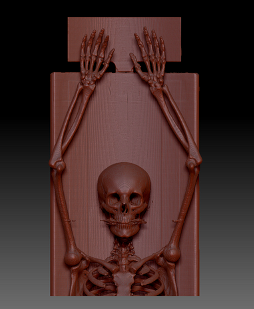
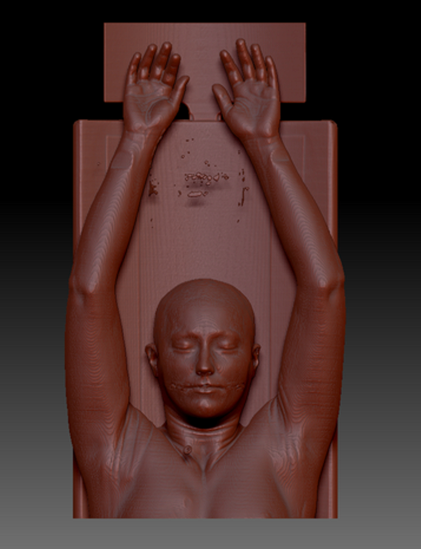
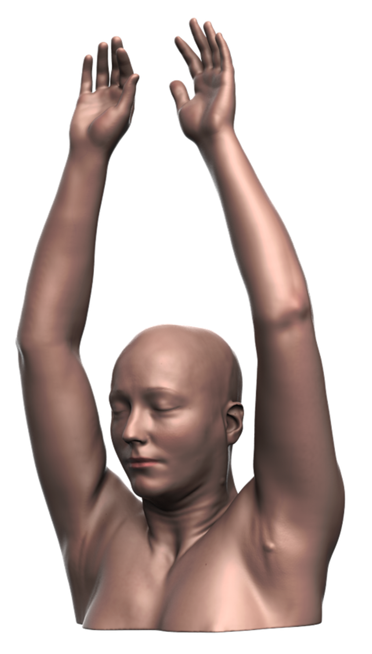
This project involved accessing real-life CT Data and reconstructing this data in ZBrush. The initial model, shown below, contained many artifacts which required removal and repairing of the affected areas. Following this, texture and colour could be added to the models, as well as lighting, in KeyShot.
I then took the final rendered image into Adobe Photoshop to create a poster designed to educate on Tularemia. This disease leads to swelling in the lymph nodes of the armpits, depicting this allowed me to make use of the raised arm position found in CT Scans.
I used Adobe Photoshop to draw the lymph nodes and the ulcers as well as Adobe Illustrator to create the insects.



Please Scroll
bottom of page